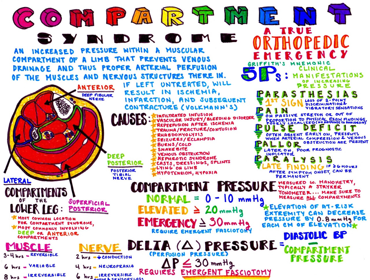
As the compartments are not designed to expand, the muscles, blood vessels and nerves within the compartment and compressed. In compartment syndrome, the pressure within a muscle compartment exceeds what is deemed normal. If we look at the lower leg, it is separated into four compartments: The muscles in each compartment have similar functions and typically work together to produce movement.

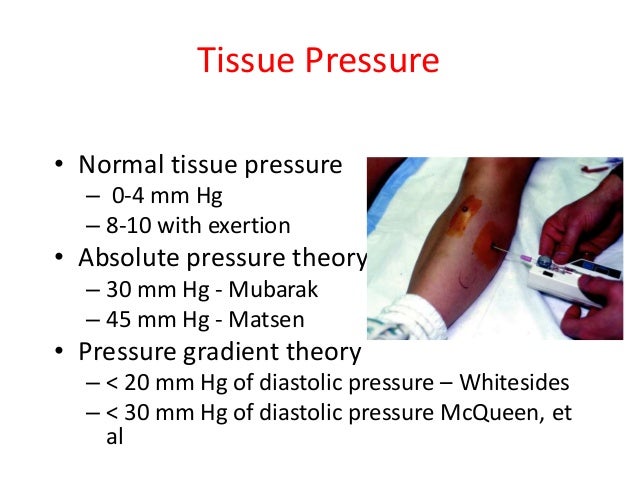
Alongside muscles, these compartments also contain nerves, blood vessels and connective tissues. This membrane allows very little stretch or expansion as it functions to keep the muscles together and hence functioning efficiently. It is important to understand that the muscles of the legs are separated into compartments that are enclosed by a connective tissue membrane (called a fascia ). While it most commonly affects the lower leg, it can also affect the arms, thighs, foot, buttocks and the abdomen. doi:10.Compartment syndrome describes the development of high pressure within a muscle compartment. Extravasation of Radiographic Contrast Material and Compartment Syndrome in the Hand: A Case Report. Belzunegui T, Louis C, Torrededia L, Oteiza J. Glossary of Terms for Musculoskeletal Radiology. MR Imaging of Compartment Syndrome of the Lower Leg: A Case Control Study. Acute Compartment Syndrome: Obtaining Diagnosis, Providing Treatment, and Minimizing Medicolegal Risk. If conservative management is unsuccessful, emergent fasciotomy is usually required for limb salvage. dressing, splint, or cast) and placing the limb at the level of the heart. Immediate management of suspected acute compartment syndrome involves relieving pressures on the compartment (e.g. Sequelae of chronic compartment syndrome include muscle atrophy, scarring, and dystrophic calcification. In myonecrosis, muscle enhancement on T1 post gadolinium sequences is absent and decreased in ischemia. The normal fascicular architecture is often lost. MRI and US may show muscle edema and swelling. In many cases, imaging may delay the diagnosis and time to surgical treatment. The utilization of imaging is generally limited 4. Etiologyīleeding in a joint or enclosed compartmentĪcute compartment syndrome is diagnosed based on clinical findings and the measurement of compartmental pressures. If the intracompartmental pressure is below the perfusion pressure, ischemia occurs which is reversible. It occurs when the interstitial pressure within the compartment exceeds the perfusion pressure of the capillary beds, causing irreversible myonecrosis due to cellular anoxia 3. This is often associated with trauma such as fractures or muscle injury.

Paralysis: this is rare and often a late findingĪcute compartment syndrome results primarily from an increase in intracompartmental pressure. Pallor: this occurs secondary to vascular insufficiency and is uncommon Paresthesia: this suggests ischemic nerve injury Often described as deep and burning in nature Patients with acute compartment syndrome often report pain in which the severity is out of proportion to the apparent injury There are five characteristic signs and symptoms ( 5 Ps) for acute compartment syndrome and they generally appear in a stepwise fashion: It is ten times more common in males and most commonly seen following tibial shaft fractures 2. Acute compartment syndrome is more common in those under 35 years of age 1.


 0 kommentar(er)
